

Standard testing of individual radiation sensitivity
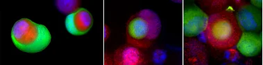
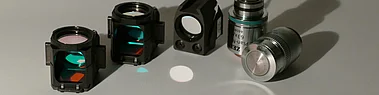
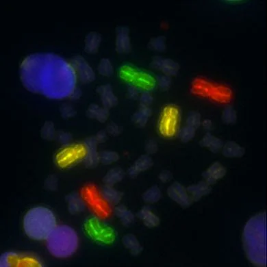
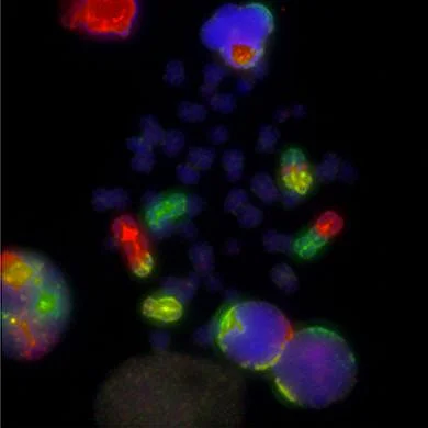
The standard testing to determine individual radiation sensitivity is the analysis of chromosomal mutations by 3-color fluorescence in situ hybridization. This requires 15ml of the patient's blood. Half of the blood is irradiated with 2 Gy of ionizing radiation (X-rays), the other half remains unirradiated. The lymphocytes in the blood, which normally do not divide, are stimulated to divide by a plant substance (lectin) and stored in an incubator at 37°C. After 48 hours, a substance is added (Colcemid) which arrests the cells during the actual cell division and thus the chromosomes can be easily assessed. Through 3-colour fluorescence hybridisation, the three largest chromosomes are provided with green, red and yellow fluorescent dyes and images of the chromosomes can be taken with an appropriate microscope. At least 100 usable images are acquired from the irradiated blood lymphocytes and 400 from the non-irradiated lymphocytes. The images are scanned for changes in the chromosomes and an average value is determined in each case. The background from the non-irradiated lymphocytes is subtracted from the value of the irradiated lymphocytes and the actual value is obtained as fractions per metaphase. If this value is higher than 0.5 breaks per metaphase there is a slightly increased radiation sensitivity. If the value is higher than 0.6 breaks per metaphase, the radiation sensitivity is already clearly increased. The examination takes between 7 and 10 days.