

Analysis of tissue sections
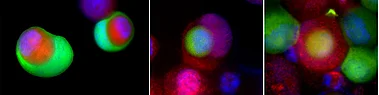
The most common way to evaluate tissue sections is to count the number of specifically stained cells and to relate the cells to the area or to the number of existing tumor cells. In addition to this procedure, we have also taken the spatial distribution into account. We investigate whether the cells are randomly distributed, or whether they deviate from a random distribution, e.g. due to cell clusters. In addition, we use double staining to examine the distances between different cell types and can thus determine the interactions between the cell types. Using both methods, we believe that we can determine the functional activity and make statements about how the tumor has suppressed an immune response.

Image analysis
For the analysis of immunohistochemically stained tissue sections we usually use double staining, e.g. cytotoxic T cells (CD8+) and regulatory T cells (FoxP3+). The entire sections are then scanned and analysed with the image analysis system Biomas, which we have developed. This enables the automatic recognition of the epithelial and stromal parts of the tumor as well as the semi-automatic recognition of the inflammatory cells. The position of the different cells in relation to each other is also calculated in this system.
